Smart Probe Tel-Aviv University: Detecting Cancer Cells May Improve Survival Rates

A new Tel Aviv University study explores a novel smart probe for image-guided surgery that may dramatically improve post-surgical outcomes for cancer patients.
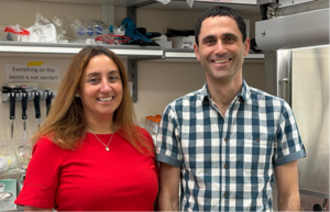
In many kinds of cancers, it is often not the primary malignant tumor, but rather metastasis — the spread of lingering cancer cells to other parts of the body — that kills patients. A multidisciplinary team led by Prof. Ronit Satchi-Fainaro of the Department of Physiology and Pharmacology at TAU’s Sackler Faculty of Medicine created a smart probe that, when injected into a patient a few hours prior to surgery to excise a primary tumor, may help surgeons pinpoint where the cancer is situated down to several cancer cells, permitting them to guarantee the removal of more cancer cells than ever before.Yana Epshtein (left) and Rachel Blau
“In cases of melanoma and breast cancer, for example, the surgeon may believe he/she has gotten everything — that he/she has excised the entire tumor and left the remaining tissue free of cancer. Even if only a few cells linger after surgery, too few or too small to be detected by MRI or CT, recurrence and metastasis may occur,” Prof. Satchi-Fainaro says. “Our new technology can guide the surgeon to completely excise the cancer.”
Making cancer cells “glow in the dark”
The new technique harnesses near-infrared technology to identify the cancer cells. “The probe is a polymer that connects to a fluorescent tag by a linker. This linker is recognized by an enzyme called cathepsin that is overproduced in many cancer types,” says Prof. Satchi-Fainaro. “Cathepsin cleaves the tag from the polymer and turns on its fluorescence at a near-infrared light.”
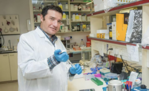
The smart probes may potentially be used to guide the surgeon in real time during tumor excision. The surgeon can also avoid cutting out any “non-glowing” healthy tissue. Prof. Ronit Satchi-Fainaro in her lab
The scientists first examined the effect of the probe in the lab on regular healthy skin and mammary tissue, and then on melanoma and breast cancer cells. They subsequently used mouse models of melanoma and breast cancer to perform routine tumor excision surgeries and smart probe-guided surgeries.
“The mice that underwent regular surgery experienced recurrence and metastasis much sooner and more often than those who underwent our smart probe-guided surgery,” says Prof. Satchi-Fainaro. “Most importantly, those which experienced the smart probe surgery survived much longer.”
Decreasing the need for additional surgery
“The probe may also reduce the need for repeated surgeries in patients with cancer cells that remain in the edges of removed tissue,” Prof. Satchi-Fainaro says. “Altogether, this may lead to the improvement of patient survival rates.”
“We are currently designing and developing additional unique polymeric Turn-ON probes for the purpose of image-guided surgery. They can be activated by additional analytes such as reactive oxygen species (ROS), which are overproduced in cancer tissues, or by using other chemiluminescent probes. We are always looking at ways to improve sensitivity and selectivity which are paramount to cancer patients’ care.”
The scientists who conducted the research for the study included Rachel Blau, Yana Epshtein and Evgeni Pisarevsky, all students in Prof. Satchi-Fainaro’s TAU lab. The research is based on long-term collaboration with Prof. Doron Shabat of TAU’s School of Chemistry, Prof. Galia Blum of the Hebrew University in Jerusalem, and clinicians Prof. Zvi Ram and Dr. Rachel Grossman of the Department of Neurosurgery at Tel Aviv Medical Center. This work was supported by the ERC Consolidator Award, the Israeli National Nanotechnology Initiative (INNI), Focal Technology Area (FTA) program: Nanomedicine for Personalized Theranostics, the Leona M. and Harry B. Helmsley Nanotechnology Research Fund, the Israel Science Foundation and the Israel Cancer Association.
Publication in Theranostics on June 21, 2018


