Un chercheur du JCT (Machon Lev, Israël) participe à une recherche révolutionnaire en imagerie cérébrale à la Cornell U. (USA)

[:fr]
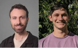
Le Dr David Sinefeld, professeur et chercheur au Jerusalem College of Technology (Machon Lev) et des scientifiques de l’Université Cornell aux USA, viennent de présenter une nouvelle méthode d’imagerie cérébrale en cartographiant le cerveau d’un poisson zèbre, offrant un aperçu sans précédent pour de futures découvertes cérébrales majeures chez l’homme.
Grâce aux laboratoires de l’université, l’équipe a utilisé des microscopes avancés afin de visionner la structure fine et l’activité d’un cerveau de poisson zèbre adulte, ce ouvre un nouvel horizon de recherche neurologique.

«Tout le monde rêvait de résoudre ce problème», se félicite le Dr Sinefeld, évoquant la difficulté à examiner avec succès des tissus cérébraux épais, en particulier à travers des écailles de poissons adultes.
Cette méthode innovante et précise fournit des photons de longueur d’onde de 1300nm à un certain point du cerveau, activant une protéine fluorescente spécifique. Le laser scanne ensuite à plusieurs reprises une certaine zone du cerveau, recueillant une image tridimensionnelle de sa structure. À ce jour, toutes les autres méthodes de recherche à l’intérieur du cerveau – comme une IRM – ne donnent pas la résolution nécessaire pour voir les neurones et la structure intérieure.
Les expériences sur le poisson zèbre représentent un champ de recherche précieux pour comprendre le cerveau humain, car les cerveaux de vertébrés sont de nature similaire. Bien que les scientifiques utilisent généralement des souris et des singes comme modèles pour le cerveau humain, le poisson zèbre représente une option tout aussi valable.
«Tous les cerveaux de vertébrés sont, à première vue, les mêmes, presque toutes les régions du cerveau existent chez presque tous les vertébrés», a déclaré Joseph Fetcho, professeur de neurobiologie et du comportement et directeur de Cornell Neurotech au College of Arts and Sciences, la revue de Cornell. «Ce n’est pas surprenant car tous, même les plus simples, doivent faire les mêmes choses pour survivre et se reproduire.»
«Nous avons utilisé une nouvelle méthode de microscopie inventée dans le laboratoire du Pr Chris Xu à la Cornell U. Dans cette méthode, nous utilisons des lasers spéciaux avec des impulsions extrêmement courtes qui interagissent avec les molécules dans le cerveau d’une manière qui permet la séparation entre cette interaction et la lumière diffusée des autres couches de tissu. Cela signifie que nous pouvons faire briller un faisceau laser à travers les écailles de poisson et voir toujours les neurones derrière eux, ce qui nous permet d’imager des neurones spécifiques au plus profond du cerveau avec une très haute résolution », a déclaré le Dr Sinefeld, qui a passé cinq ans en tant que postdoctorant chercheur au centre Cornell Neurotech.
«Cette méthode ouvre un nouvel horizon pour la recherche sur le cerveau animal. Nous pouvons maintenant mieux voir comment fonctionne le cerveau », a ajouté le Dr Sinefeld. «Cette recherche nous permet de surveiller un cerveau de poisson zèbre complet au fil du temps. Par exemple, après avoir appliqué cet outil à des poissons conçus pour souffrir de certains troubles cérébraux, les images peuvent alors déchiffrer comment le cerveau change à mesure que le poisson mûrit. De même, les images peuvent également voir comment les poissons réagissent au traitement au fil du temps et peuvent avoir des implications dramatiques dans la façon dont nous comprenons les fonctions cérébrales et leurs troubles ».
Le Dr Sinefeld veut poursuivre ses efforts dans le domaine de la microscopie au JCT et recherche actuellement des subventions et des financements pour construire un nouveau laboratoire dédié à cette discipline à l’école. «Cela pourrait changer la donne dans le domaine des neurosciences. Je suis ravi d’avoir l’opportunité d’établir ces nouvelles méthodes en Israël et en particulier au JCT », a-t-il déclaré.
« Créé en 1969, le Jerusalem College of Technology est l’une des institutions universitaires les plus prestigieuses d’Israël, en science et technologie. Le JCT fournit des diplômés professionnels hautement qualifiés à l’industrie de la haute technologie en Israël et dans le monde. C’est la seule institution d’enseignement supérieur engagée à fournir une éducation universitaire du plus haut niveau à diverses catégories de la société israélienne qui n’auraient pas pu accéder à ces disciplines. Le JCT propose des programmes exclusifs développés spécifiquement pour les hommes et les femmes Haredim (ultra-orthodoxes) et pour d’autres catégories de la population », précise Noah Dana-Picard, directeur de la Chaire Mathématiques, Education et Torah au JCT.
Traduit et adapté par Esther Amar pour Israël Science Info
[:en]
Jerusalem College of Technology lecturer and researcher Dr. David Sinefeld along with his Cornell University counterparts reveal new method for brain imaging in zebrafish, potentially providing great insight into future brain discoveries.
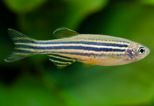
Israeli researcher and Jerusalem College of Technology professor Dr. David Sinefeld and a group of interdisciplinary scientists at Cornell University have made a new breakthrough in brain imaging thanks to its mapping of a zebrafish’s brain.
The team, working in the university’s labs, used advanced microscopy methods in order to image the fine structure and activity of an adult zebrafish brain which resulted in opening a new horizon of neurological research.

“This is a problem that everyone dreams of solving”, Dr. Sinefeld said, referring to the difficulty in successfully examining thick brain tissue, especially through adult fish scales.
Experimenting on zebrafish is a useful stepping-stone to understanding the human brain because all vertebrate brains are similar in nature. Although scientists usually use mice and monkeys as models for the human brain, zebrafish are another viable option.
“All vertebrate brains are, to a first approximation, the same, with nearly all brain regions [present] in nearly every vertebrate,” said Joseph Fetcho, professor of neurobiology and behavior and director of Cornell Neurotech in the College of Arts and Sciences told the Cornell Chronicle. “This is not surprising because they all, even the simplest ones, have to do the same things to survive and reproduce.”
 “In order to solve this problem, we used a novel microscopy method which was invented in Prof. Chris Xu lab [at Cornell]. In this method, we use special lasers with extremely short pulses which interact with the molecules in the brain in a way that allows separation between this interaction and the scattered light from other tissue layers. This means that we can shine a laser beam through fish scales, and still see the neurons behind them, allowing us to image specific neurons deep in the brain with very high resolution”, said Dr. Sinefeld, who spent five years working as a postdoctoral researcher Mong Fellow at Cornell Neurotech center.
The innovative, precise method delivers 1300nm wavelength photons to a certain point in the brain, activating a specific fluorescent protein. The laser then repeatedly scans a certain section of the brain, garnering a three-dimensional image of its structure. To date, all other methods of looking inside the brain – like an MRI – don’t yield the resolution needed to see the neurons and structure inside.
“This method opens a new horizon for animal brain research. We can now see better how the brain works”, Dr. Sinefeld added. “This research allows us to monitor a full zebrafish brain over time. For instance, after applying this tool to fish engineered to have certain brain disorders the images can then decipher how the brain changes as the fish mature. Likewise, the images can then also see how the fish respond to treatment over time and can lead to dramatic implications in how we understand brain functions and their disorders.”
Dr. Sinefeld hopes to continue his efforts in the field of microscopy at the Jerusalem College of Technology (JCT) and is in the process of applying for grants and funding to build a new lab dedicated to this discipline at the school.
“This could be a game-changer in the field of neuroscience. I am excited for the opportunity to establish these novel methods In Israel and specifically in JCT”, he said.
Established in 1969, the Jerusalem College of Technology (JCT) is one of Israel’s most prestigious and unique academic institutions with a focus on science and technology. JCT supplies highly skilled, professional graduates to Israel’s and the world’s high-tech industry. In addition, we are the only institution of higher learning committed to providing the highest quality academic education to diverse segments of Israeli society who would not otherwise have had the opportunity to enter these fields. JCT offers exclusive programs developed specifically for Haredi (ultra-Orthodox) men and women and for other demographic groups.
[:]

